Yahya Chaudhry
Harvard Staff Writer
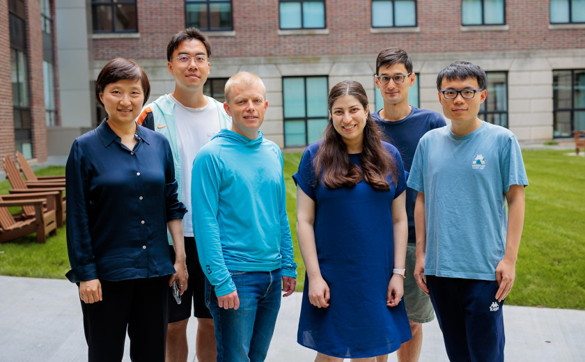
The technique overcomes a key limitation of existing imaging tools, letting researchers chart nearly 10,000 RNA isoforms across mouse brain tissue
Harvard Staff Writer
Harvard researchers have unveiled a new way to map the molecular activity in complex tissues that can reveal which specific RNA variants genes produce in every cell and every region of the tissue.
The technology, described in a new paper in Cell, could reshape how scientists investigate tissue function and disease. The work comes from the lab of Xiaowei Zhuang, David B. Arnold Jr. Professor of Science in the Department of Chemistry and Chemical Biology. Her group is known for pushing microscopy beyond traditional limits and previously developed MERFISH, a genome-scale imaging method that can measure thousands of RNA molecules in intact tissues with extremely fine spatial detail. The researchers demonstrated this technology in the brain, and it may extend to cancers and immune disorders as well.
“Imaging-based spatial transcriptomics gives you a very high spatial resolution,” Zhuang said. “You can see individual cells. Not only that — but you can also get subcellular resolution.”
Spatial transcriptomics, which map gene expression directly within the physical architecture of a tissue, have largely followed two paths. Imaging-based methods like MERFISH deliver high spatial resolution and powerful detection but typically require researchers to choose a subset of genes. Sequencing-based approaches sample more broadly from the transcriptome — all genes in a sample — but lack single‑cell or subcellular resolution and struggle with low detection.
Researchers have long aimed to have it both ways: whole‑transcriptome coverage and single‑cell, spatial resolution with high detection. On paper, MERFISH’s combinatorial barcoding could scale to tens of thousands of RNA targets to cover the whole transcriptome. In practice, a more basic constraint got in the way: transcript length — imaging and identifying short RNA molecules — is very challenging.
“In MERFISH, we need to detect RNA at the single-molecule level, and usually one probe is not enough to detect that molecule. We need to tile dozens of probes per molecule to get a good signal,” Zhuang, who also holds the title of Howard Hughes Medical Institute investigator, said, “but some transcripts are very short and cannot accommodate so many probes.”
That limitation is especially severe for short regulatory RNAs and RNA isoforms — variants of the same gene that differ only in small segments. Those differences are crucial in the brain, where alternative splicing and other transcript variations help define cell types and circuit properties, but they are difficult to see with existing tools.
To address this, Limor Cohen, a postdoctoral scholar in the Zhuang lab and lead author of the paper, led the development of an in situ amplification strategy called RT&T-AMP (Reverse Transcription and Transcription-Mediated Amplification). First, each RNA molecule in a fixed tissue slice is converted into complementary DNA, then used as a template to synthesize many RNA amplicons at that same location. The effect is to turn every original transcript into a localized cluster of many identical RNA copies that can be detected with far fewer probes.
The amplified RNAs are then read out using MERFISH imaging.
With this RT&T‑AMP/MERFISH platform, the researchers imaged about 33,000 distinct RNA sequences in mouse brain tissue — roughly 23,000 genes and 10,000 annotated isoforms — at single‑cell resolution. The data allowed them to build molecular maps of major brain regions, identify cell types and states, and chart ligand-receptor-mediated cell‑cell communications.
Crucially, they could also see which isoforms were used in which cell type and where — something that has until now been largely inaccessible at scale.
“Going into the project, we didn’t really know what to expect because there wasn’t that much knowledge about cell-type- and brain-region-specific isoform expression,” said Cohen. “We were encouraged to see all these widespread isoform changes in different brain regions and different cell types, and I think it’s an important layer of biology that’s currently missing in the field.”
Some brain regions show particularly rich isoform specificity. For example, the team found isoforms uniquely enriched in subregions of the hippocampus, the area central to learning and memory.
While the paper focused on isoforms, Zhuang sees broader potential. The approach, she said, has “power in terms of measuring transcriptome-wide information, including not only messenger RNAs and their isoforms, but also regulatory non-coding RNAs, including potentially microRNAs and other small RNAs.”
The scale of information emerging from this single experiment is enormous. Applications may extend to cancer and immune disorders.
Going forward, Zhuang hopes to extend applications of the method beyond the mouse brain to human tissue and disease models, and to combine it with genetic perturbations — a laboratory technique used to intentionally alter gene activity — and AI-based modeling. By layering these spatial maps with functional experiments, they hope to move from describing RNA patterns to explaining how variations in genes and isoforms impact health and disease.

Two Harvard scientists describe a way to engineer the evolutionary landscape that viruses experience, pushing them away from dangerous variants and into evolutionary dead ends.

Amid the rhythms of the back-to-campus bustle, new faculty find themselves experiencing the same first-day excitement — and jitters. We dropped in on three, on the first day of their first classes.

As the sun sinks below the horizon, two scientists perch quietly beside a darkening field and wait for flashes of light. They have traveled far outside the city in search of research sites, because they study a group of creatures that have been driven to the margins of human settlements: fireflies.