Eric Moskowitz
Harvard Staff Writer
Harvard Staff Writer
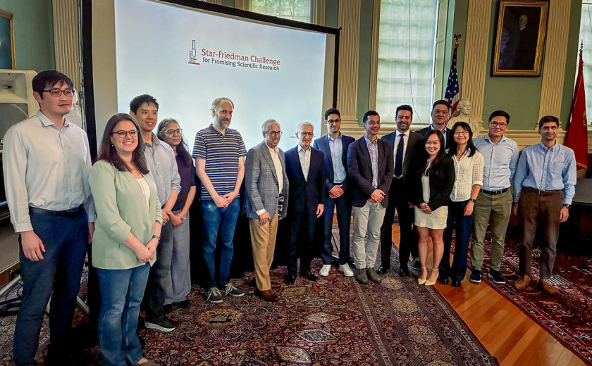
Pictured above, left to right: Kiyoul Yang, Assistant Professor, Harvard John A. Paulson School of Engineering and Applied Sciences (SEAS); Giulia Semeghini, Assistant Professor of Applied Physics, SEAS; Xing Fan, Assistant Professor of Physics; Victoria D’Souza, Professor of Molecular and Cellular Biology; Doeke Hekstra, Associate Professor of Molecular and Cellular Biology, Associate Professor of Applied Physics, SEAS; donor James A. Star ’83; donor Josh Friedman ’76, M.B.A. ’80, J.D. ’82; Mahmoud Mikdar (research associate representing Professor Manoj Duraisingh, John LaPorte Given Professor of Immunology and Infectious Diseases, Harvard T.H. Chan School of Public Health); Ahmad “Mo” Khalil, Hok Lam and Kathleen Kam Wong Professor of Bioengineering, SEAS, Professor of Molecular and Cellular Biology; Maxim Prigozhin, Assistant Professor of Molecular and Cellular Biology and of Applied Physics, SEAS; Kathy Liu (front; graduate student representing Joanna Aizenberg, Amy Smith Berylson Professor of Materials Science, SEAS, Professor of Chemistry and Chemical Biology); Donhee Ham John A. and Elizabeth S. Armstrong Professor of Engineering and Applied Sciences, SEAS (rear); Chenghua Gu, Professor of Neurobiology, Harvard Medical School; Quan Lu, Cecil K. and Philip Drinker Professor of Environmental Physiology, Harvard T.H. Chan School of Public Health; selection committee chair Michael Desai.
The 2026 Star-Friedman Challenge grants will support seven “high risk, high reward” projects pursued by Harvard researchers, including engineering red blood cells to patrol the body as disease detectors, designing microchip-sized lasers for molecular experiments and quantum computing, and developing tactile-sensing “skins” to help robots achieve more human-like touch in assisting with senior care, surgery, or disaster recovery.
“I’m really struck by the ambition and range of the work,” said Hopi Hoekstra, Edgerley Family Dean of the Faculty of Arts and Sciences, at a University Hall celebration for the winners and their projects. “It really reflects the extraordinary creativity of our faculty and the exciting possibilities that emerge when researchers are given the freedom and support, encouragement, and opportunity to really follow and pursue their boldest ideas.”
The endowed program, which provides seed funding of $80,000 to $150,000 for faculty conducting investigations in the life, physical, and social sciences, received 88 applications this year. This record number reflects not only the dynamism and ambition of Harvard researchers, but also their collective uncertainty around federal funding.
Even in the best of times, federal funding more often flows to established projects making incremental advances, said Michael Desai, the selection committee chair. “That’s why programs like this Star-Friedman Challenge are really so important and offer such great opportunities for us to try out new ideas, to move into new directions, and to shift our research programs into exciting new areas,” said Desai, the Fisher Professor of Natural History in the Department of Organismic and Evolutionary Biology.
Officially the Star-Friedman Challenge for Promising Scientific Research, the program was established in 2013 by a gift from James A. Star ’83 and expanded five years later with support from Josh Friedman ’76, M.B.A. ’80, J.D. ’82, and Beth Friedman. It encourages investigators to collaborate across disciplines and branch into new directions.
As a life sciences scholar herself, Hoekstra was heartened to see so many early-career faculty among this year’s recipients. “I know firsthand how that early investment can really impact both the direction and trajectory of one’s scientific career,” she said, at the June 11 ceremony. “Some of the most important discoveries begin not with certainty but with a promising idea or … the opportunity to take intellectual risks.”
Joanna Aizenberg
Amy Smith Berylson Professor of Materials Science, Harvard John A. Paulson School of Engineering and Applied Sciences
Professor of Chemistry and Chemical Biology
Each human fingertip contains thousands of “mechanoreceptors” with the ability to sense force, texture, and movement in milliseconds. By comparison, advanced robots today with touch capabilities fit, at most, a few hundred sensing elements in the same area. This makes them good at handling predictable objects but still clumsy when it comes to grasping complex or deformable items such as laundry, bags of fruit, or slippery material. That’s because those robotic systems rely on arrays of discrete electronic sensing elements connected by complex wiring and readout circuitry, which don’t also lend themselves well to scaling up for robots with large, curved, or varying sizes and shapes. Aizenberg’s lab proposes a new kind of micro-structured “smart skin” that would do away with the wiring and electronics by changing color across an array of tiny pixels — optical signals that could be read by cameras and computer vision to enable safer and more sensitive dexterity in robotic applications such as elder care, surgery, and delicate manufacturing.
Ahmad “Mo” Khalil
Hok Lam and Kathleen Kam Wong Professor of Bioengineering, SEAS
Professor of Molecular and Cellular Biology
Manoj Duraisingh
John LaPorte Given Professor of Immunology and Infectious Diseases, Harvard T.H. Chan School of Public Health
The human body’s 25 trillion red blood cells deliver oxygen from the lungs to tissues throughout the body, while continually being replenished during their 120-day circulatory span. But what if these cells could be harnessed not just for delivery but for disease detection, too? Duraisingh’s lab studies red blood cell biology as it relates to malaria, to understand host-parasite interactions and to inform vaccine and drug development for a mosquito-borne disease that claims half a million lives globally each year. Khalil, a new professor on the other side of campus, is a synthetic biologist who works on reprogramming cells to treat disease, such as CAR T-cell therapy that genetically engineers white blood cells to target certain blood cancers. Their collaboration aims to “combine engineered erythroid systems with synthetic sensing receptors to create PATROL-RBCs” — in other words, to program red blood cells to switch on a detectable signal if they encounter diseases while circulating through the body, enabling earlier and more accurate diagnoses as well as the potential for targeted treatments.
Donhee Ham
John A. and Elizabeth S. Armstrong Professor of Engineering and Applied Sciences, SEAS
Back in 1949, Donald Hebb theorized that when neurons in the brain repeatedly fire together, their synapses — the microscopic gaps where neurons, or nerve cells, meet and transmit electrical impulses — strengthen. This “fire together, wire together” postulate has become foundational. But even after three-quarters of a century, no one has been able to watch this rewiring unfold in detail across a living neural network, with a trade-off between detailed observation inside a few neurons or less sensitive observation of a wider network. Ham’s lab has already developed a breakthrough device known as the intracellular microelectrode array (iMEA), culturing rat cortical neurons on a specially designed silicon chip outfitted with 4,000 microscopic electrodes. In a demonstration, iMEA mapped over 70,000 plausible synaptic connections while classifying their strength and type. With that “scale-detail divide broken,” Ham and his team plan to use the Star-Friedman grant to conduct a continuous study of how brain cells rewire themselves as a neural network learns, with potential applications for computing and disease treatment.
Doeke Hekstra
Associate Professor of Molecular and Cellular Biology
Associate Professor of Applied Physics, SEAS
Victoria D’Souza
Professor of Molecular and Cellular Biology
The shapes and structures of biology’s most important proteins — including antibodies, viral-replication machinery, and the ones that transcribe DNA into mRNA — can be glimpsed at the atomic level using two different primary methods. They can be crystallized and studied using X-ray crystallography or frozen and examined using cryogenic electron microscopy. Both methods present challenges, because freezing and crystallizing can affect the shape of a protein — but also because the proteins themselves regularly shift and change their shapes in their natural environments, in ways that freezing and crystallizing fail to capture. Hekstra and D’Souza instead propose examining proteins in solutions that mimic their physiological conditions, while using electric-field-oriented wide-angle X-ray scattering. By giving the proteins a nanosecond pulse of electricity, they intend to orient them into a position that allows for a better snapshot, without causing heating or electrochemical changes. Hekstra said the idea occurred to him while listening to a recent academic talk on RNA dynamics. He considered the historic Photo 51 — which achieved a sharp image of DNA through X-ray diffraction all the way back in 1952, even as proteins have remained hard to photograph. Hekstra thought that it might be easier if he could align protein molecules as neatly as DNA, hypothesizing it could be done with brief pulses. His proposal coupled his experience with X-ray diffraction and time-resolved experiments with electrical pulses with D’Souza’s work using X-ray scattering to study the biochemistry and biology of HIV-1. Understanding the real shape of protein molecules could have a range of benefits, including validating drug targets.
Maxim Prigozhin
Assistant Professor of Molecular and Cellular Biology and of Applied Physics, SEAS
Prigozhin wants to understand the varying conformations — meaning the shapes and structures — of individual proteins beyond the limited glimpses afforded by X-ray crystallography and cryo-electron microscopy. But where Hekstra and D’Souza propose aligning the proteins with electrical pulses before X-raying them, Prigozhin will apply pressure to excite them before trapping them with flash freezing and examining them with cryo-electron microscopy. He intends to build an instrument that will gently squeeze the proteins to reveal rare, hidden shapes that standard freezing techniques miss. Discovering these shapes and states could reveal drug-binding pockets while opening up new targets for medicines.
Quan Lu
Cecil K. and Philip Drinker Professor of Environmental Physiology, Harvard T.H. Chan School of Public Health
Chenghua Gu
Professor of Neurobiology, Harvard Medical School
A thin layer of cells known as the blood-brain barrier serves as a gatekeeper between the circulatory and central nervous systems, shielding the brain from toxins and infection. Essential for survival, that same barrier also blocks nearly all large and biologic therapeutic drugs — meaning many otherwise-promising treatments for neurodevelopmental disorders, neurodegenerative diseases, and brain tumors can’t reach their targets. To bypass it, scientists have developed two primary methods: disrupting the barrier with ultrasound or osmotic shock, and loading the therapeutics onto molecules that act as “brain shuttles” by tricking receptors on the barrier to let them in. But disrupting the barrier is invasive and lacks molecular precision, and the current shuttle method is not “brain-specific,” leading the drug to accumulate in other tissues, which can cause severe side effects. Lu and Gu propose engineering a type of vesicles known as ARMMs into a programmable, precision delivery system that could cross the blood-brain barrier, carry therapies to the brain, and deliver them specifically to chosen cell types, which could potentially unlock gene editing, mRNA medicines, and other biologic treatments for brain disorders currently considered untreatable.
Kiyoul Yang
Assistant Professor of Electrical Engineering, SEAS
Xing Fan
Assistant Professor of Physics
Giulia Semeghini
Assistant Professor of Applied Physics, SEAS
Researchers who work with molecules and atoms to advance the understanding of quantum physics and enable progress on technologies such as quantum computing rely heavily on lasers and optical equipment — mirrors, lenses, beamsplitters, and more — to manipulate and image them. Those lasers require precision tuning and take up a lot of space. A simple misstep in a lab or a powerful storm outside can cause a painful setback, a problem that Fan and Semeghini commiserated about as office neighbors in the Goel Quantum Science and Engineering Building. But what if they could do for lasers and optics what microprocessors did for computing, shrinking the computer from the size of a room to a tiny device? Enter Yang — an expert in developing chip-based photonic systems for computing, sensing, communications, and precision measurement — for a cross-disciplinary collaboration aimed at designing chip-based light circuits that could reliably deliver and control laser beams for atoms and molecules, potentially making next-generation quantum computers and sensors more robust and scalable.
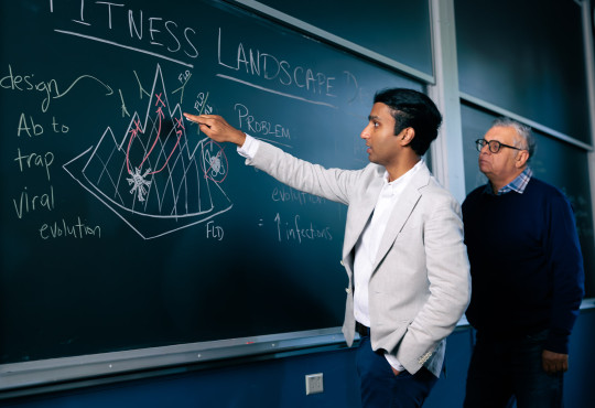
Two Harvard scientists describe a way to engineer the evolutionary landscape that viruses experience, pushing them away from dangerous variants and into evolutionary dead ends.

Amid the rhythms of the back-to-campus bustle, new faculty find themselves experiencing the same first-day excitement — and jitters. We dropped in on three, on the first day of their first classes.

As the sun sinks below the horizon, two scientists perch quietly beside a darkening field and wait for flashes of light. They have traveled far outside the city in search of research sites, because they study a group of creatures that have been driven to the margins of human settlements: fireflies.