Wendy Heywood
Harvard Correspondent
OEB researchers discover “cautionary tale” about fish brains
Harvard Correspondent

For every two living vertebrates on Earth, one is a fish — 95% of which are ray-finned fish such as salmon, tuna, goldfish, and trout. Roughly 35,000 species comprise ray-finned fish, and they thrive in environments ranging from deep-sea trenches to alpine Himalayan rivers, Amazonian floodplains, and desert ponds. Despite their abundance, little is known about the diversity of their brains.
“How can we pretend that we understand how evolution, and especially brain evolution, works if we know almost nothing about half of the vertebrates?” asked Rodrigo Figueroa, postdoctoral researcher in the Department of Organismic and Evolutionary Biology (OEB) and biodiversity fellow at Harvard's Museum of Comparative Zoology (MCZ).
A new study published in Proceedings of the Royal Society B aims to change that.
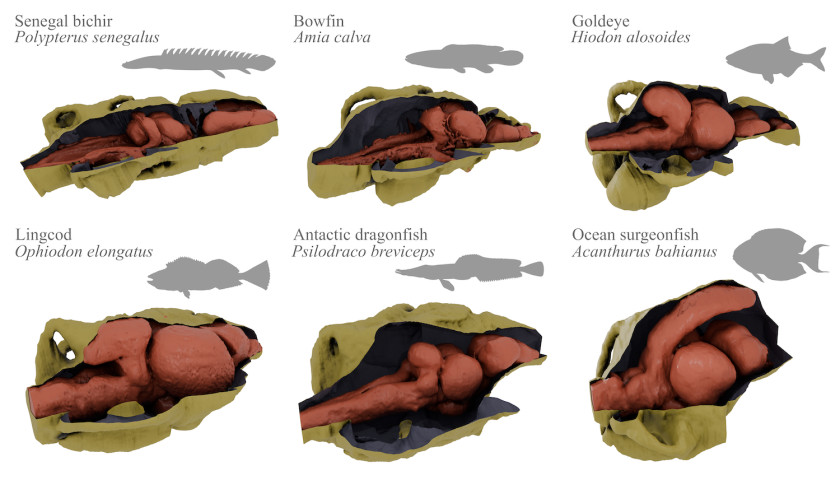
The researchers, led by Figueroa, used a special CT scanning technique to peer inside the heads of 87 ray-finned species across more than 70 families to map their internal structures in three-dimensional detail. The findings revealed a surprising neurological landscape where brain size and shape, as well as endocasts — the mold of the internal skull cavity — vary wildly.
The project grew from Figueroa’s Ph.D. research at the University of Michigan on extinct ray-finned fishes. That work showcasing an ancient, fossilized fish brain led him to discover the lack of descriptions of living fishes’ brains; current understanding has been confined to a few model organisms like the zebrafish and sporadic descriptions of oddball species. To better understand the 300-million-year-old fossil, Figueroa embarked on a five-year mission to CT scan living species of ray-finned fishes.
The most striking finding was a ratio comparing brain volume to intracranial space. In most vertebrates, like reptiles and mammals, the brain fills the cranial cavity snugly, making them predictably comparable. In ray-finned fishes, most cluster around 40 to 50 percent, and some possess brains occupying less than 5 percent of their intracranial space.
“If you CT scanned a mammal skull and made a replica of the brain by filling in the empty space inside, it would look very similar to the actual brain; there is a very tight fit between the brain and skull,” said co-author Stephanie Pierce, professor in OEB and curator of vertebrate paleontology in the MCZ. “But, with these fishes, there’s small brains, big brains, smooth brains, convoluted brains; it’s just astonishing the amount of diversity these animals are showing.”
Environmental factors appear to drive this variation, with statistical analysis linking the ratio to water depth: deep-sea fishes, whether on the ocean floor or in the open water column, tend toward smaller brains relative to skull size. The extra space is not wasted; it contains cerebrospinal fluid, blood vessels, and in some cases, specialized organs managing blood production and immune response — much like the spleen and bone marrow in humans.
“Many things can explain why it would be beneficial to have a tiny brain and a big head,” said Figueroa. “The meningeal tissue surrounding the tiny brain acts as a protective bumper, keeping it safe from impact or variations in overwhelming pressure.”
The study also tracked how brain size changes with growth. In bowfin fish, for example, the brain drops from near-total skull occupancy in hatchlings to 20-30 percent in mature adults. This trend is mild in humans and birds, but extreme in certain fish lineages. In the coelacanth, a famous living fossil, the brain fills nearly the entire cavity in youth before shrinking to a staggering 4 percent in adulthood.
Pierce called the study a “cautionary tale.” “For decades, researchers assumed a fossil’s endocast shape directly reflected its brain morphology, and while that’s a pretty good assumption for most vertebrates, it isn’t for fish,” Pierce said. “By comparing endocasts to brains, our findings show that the two can evolve on completely different trajectories.”
Ray-finned fishes’ evolutionary history and drivers of success are significant for evolutionary biology and neuroscience.
“Modern neuroscience often focuses on mapping the ‘connectomes’ of a few specific species,” Figueroa noted, “yet this study shows that such a focus provides only a narrow view of what has actually evolved over millions of years — and it raises more questions about whether the flexible, diverse brain shapes of these fishes caused their global success, or whether their success in varied environments forced their brains to adapt in such unique ways.”
The study, which relied heavily on museum collections, provides a new map for studying the nervous system’s evolution and function, reminding scientists that the highly standardized brains of mammals and birds are unique.
“This study represents our first steps,” Pierce said. “It’s giving us a quick snapshot of what the major trends are, and where we go next.”
With 87 species down and roughly 35,000 to go, Figueroa acknowledges the scale of what remains to be studied. “Maybe ten of my lifetimes and all my students in the future,” he said, laughing.

Why do so many people feel lonely in a world designed for connection? As Ian Corbin sees it, Americans are experiencing a crisis of belonging, which he attributes to some fundamental misunderstandings about what humans need to flourish.


Weiss, professor of the classics and an expert on ancient Greek culture plans, to prioritize the expansion of educational resources.